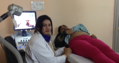
Role of Ultrasound in Pregnancy
It uses high frequency sound waves to build a picture of baby in the womb.
Safety: Usage for diagnostic purpose has no known side effects on mothers and babies.
Timing:It can be carried out at any stage of pregnancy.During early pregnancy it will be required to hold urine to visualise the baby.Transvaginal ultrasound is safe and has gives better image help in increasing the chances of correct diagnosis results in early pregnancy.

INFORMATION WITH ADVANCING PREGNANCY:
First Trimester[1] (upto 12weeks) :
The initial scan is usually done during 6-8 weeks of pregnancy in order to determine the estimated due date of delivery. It also provides first glimpse of baby.it confirms viability by documenting the feral heart activity.Done on full bladder or Transvaginal .
Second[1] Trimester[2] (13 to 28weeks) :
the second and very important scan should ideally be done at 19-20 weeks for detailed study of fetal structure and to find out if there is any Anatoly. It takes longer time to conduct the scan (,around 20 to 45 minutes. ). At this time the length measurement of cervix is also important.